
by Dena Denali | Dec 3, 2025 | Ask The Expert
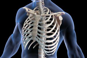
Many people come into my practice describing what they believe is a “pinched nerve,” a catch-all slang term used to describe nerve pressure without understanding the actual mechanism of injury. Often, they picture bones twisted out of place and physically shearing a nerve between them. While this can occur, it is actually one of the least common ways nerves become irritated.
More often, nerve compression and nerve entrapment syndromes arise from soft tissues—muscles, fascia, ligaments, scar tissue, or surrounding anatomical tunnels—that have become tight, inflamed, or dysfunctional.
Understanding these conditions matters, because nerve symptoms can feel alarming: burning, numbness, tingling, weakness, deep aching, or sharp electrical “zings” that travel down an arm or leg. And because nerves travel long pathways from the spine into the limbs, the true source of irritation is frequently misdiagnosed or misunderstood.
Nerve Compression vs. Nerve Entrapment: What’s the Difference?
NERVE COMPRESSION occurs when a nerve is irritated by pressure from surrounding soft tissues—often muscle tension, fascial adhesions, swollen tissues, or joint inflammation. In these cases, the soft tissues themselves are responsible for the compression.
NERVE ENTRAPMENT is a more specific form of compression where the nerve becomes irritated within a defined anatomical tunnel or passageway. Entrapments can be caused by tight muscles, scar tissue after injury or surgery, repetitive strain, localized inflammation, or structural narrowing of these bony pathways.
Examples include Carpal Tunnel Syndrome, Cubital Tunnel Syndrome, Thoracic Outlet Syndrome, and Tarsal Tunnel Syndrome.
Common Misdiagnoses & Why They Happen
Nerve issues often mimic other conditions, and it’s easy for both clients and clinicians to chase symptoms instead of identifying the source. Common misdiagnoses include:
-
Carpal Tunnel Syndrome misdiagnosed when the true issue is cervical nerve root compression, thoracic outlet syndrome, pronator teres entrapment, or shoulder dysfunction.
-
Sciatica misdiagnosed when symptoms arise from weak or misfiring glutes, sacroiliac joint dysfunction, piriformis syndrome, deep hip rotator tension, or lumbar facet irritation.
-
Rotator cuff pain misdiagnosed when nerve entrapment in the neck, pec minor, or upper ribs is actually referring pain into the shoulder.
-
Tingling in hands or feet mistaken for circulation issues when the true culprit is a nerve pathway obstruction.
Misdiagnosis occurs because nerve pain travels—it radiates, refers, jumps, and often appears far away from the actual compression.
This is where a thorough clinical assessment becomes essential.
It’s Not Usually Bones “Pinching” Nerves
While vertebral compression can occur with disc herniations or severe degeneration, most nerve irritations arise from soft tissues, not bones. Most nerves become irritated because they cannot glide, stretch, or move normally—not because a bone has sheared them.
Common causes include:
1. Muscle Tightness or Hypertonicity
When a muscle stays tight for long periods due to stress, posture, or repetitive work, it can close down space around a nerve. Even though muscle is soft tissue, sustained tension can create enough pressure to overstimulate a nerve.
2. Myofascial Restrictions
Adhesions and scar tissue form as disorganized webs—much like a bird’s nest or spider web—to stabilize an injury. As they spread, these tissues can bind or tether a nerve, limiting its ability to glide smoothly.
3. Inflammation & Compromised Anatomical Tunnels
Chronic inflammation from overuse, repetitive strain, autoimmune processes, or surgery can crowd narrow nerve pathways. Rigid tunnels, such as the carpal tunnel or the thoracic outlet space, cannot expand to accommodate swelling. The increased fluid behaves like a soft-tissue/bony vise, putting direct pressure on the nerve.
4. Postural and Biomechanical Factors
Your body, much like a vehicle, is meant to function in a relatively “square” and aligned structure. You can still drive a car that needs an alignment, but over time it wears out ball joints, tie-rod ends, and tires.
Similarly, forward-head posture, prolonged sitting, overhead work, gait abnormalities, or athletic strain can subtly compress nerves over time. These activities aren’t inherently harmful, but balanced mobility, stretching, and strengthening are essential to prevent nerve irritation.
5. Systemic Factors
Systemic conditions can make nerves more reactive and less tolerant of pressure, even when mechanical compression is mild. Fluid retention, hormonal fluctuations, diabetes, and inflammatory disorders can increase swelling, change connective tissue tension, or reduce circulation—making nerve symptoms feel more intense. Understanding these influences helps guide both treatment planning and realistic recovery expectations.
The Symptom Picture
Short-term symptoms of nerve irritation may include burning, tingling, or “pins and needles,” along with sharp, shooting, or electric-type pains. Clients may experience limb weakness or heaviness, increased muscle tightness or guarding, and difficulty gripping or weight-bearing.
If left untreated, long-term complications can develop: persistent weakness, muscle atrophy, loss of sensation, chronic pain cycles, and altered movement patterns that create secondary injuries.
Nerves are resilient—but they require adequate space, mobility, and circulation to function properly.
The Road to Resolution
At Body Kneads Integrative Healing, nerve-related cases are assessed using orthopaedic testing, anatomical pattern recognition, and integrative manual therapy. Because symptoms alone rarely reveal the true source, the goal is to identify where along the pathway the nerve is compromised.
A systematic approach helps determine whether symptoms stem from the nerve root, a peripheral entrapment, postural strain, myofascial restrictions, or—most commonly—a combination. I begin with special orthopaedic tests that produce predictable, repeatable responses, followed by palpation, movement analysis, and postural evaluation. A rule-out process differentiates muscular referral pain from true neural irritation.
Goals of Treatment
Treatment combines multiple manual therapy techniques along with Dolphin MPS microcurrent when clinically appropriate. The aim is not to chase symptoms, but to correct the underlying contributors. Treatment goals include:
-
Reducing tension around nerve pathways
-
Improving nerve mobility and glide
-
Decreasing inflammation
-
Improving posture and biomechanics
-
Restoring normal muscle recruitment patterns
-
Breaking down adhesions
-
Supporting long-term neuromuscular health
Addressing both the symptom and the structure reduces irritation and prevents recurrence.
What Recovery Looks Like
Recovery depends on how long the nerve has been irritated, the root cause(s) identified, how much postural or movement correction is needed, and patient commitment to home care. A newer problem in a motivated patient can improve quickly—often within a few sessions. Long-standing conditions may require more time due to compensatory changes.
Home care is always part of a successful plan. This may include stretching overactive muscles, strengthening underused muscles, nerve-flossing techniques, ergonomic adjustments, and guided return to normal activities or sport.
Nerves heal—but they heal best when the tissues around them move well, glide well, and are not overly tight or inflamed.
Final Thoughts
Nerve compression and nerve entrapment syndromes are incredibly common—and frequently misunderstood. They are not always the result of bones pinching nerves. More often, they occur because soft tissues surrounding the nerve have become tight, irritated, or inflamed.
Through thorough assessment and integrative hands-on therapy, we can identify the true source of irritation and create a treatment plan that restores mobility, reduces pain, and supports long-term nerve health.
If you’re experiencing numbness, tingling, weakness, or nerve-related discomfort, you don’t need to guess what’s going on. With proper assessment and targeted treatment, meaningful improvement is absolutely possible.

by Dena Denali | Nov 4, 2025 | Ask The Expert
Why They Get Confused & How to Tell the Difference
When hand pain, numbness, or tingling begins, many people assume Carpal Tunnel Syndrome (CTS) is to blame. Yet a large percentage of cases diagnosed as “carpal tunnel” actually originate much higher in the body—often in the neck, shoulder, or thorax.
A common imposter? Thoracic Outlet Syndrome (TOS).
Both conditions can create numbness, tingling, and weakness in the hand… but they are not the same problem, and they require different treatment strategies.
This is where skilled clinical assessment matters. At Body Kneads Integrative Healing, we use advanced orthopaedic testing, anatomical pattern recognition, and integrative manual therapy—including microcurrent through Dolphin MPS—to determine the true source of symptoms and treat the root cause, not just the site of pain.
Carpal Tunnel Syndrome: A Quick Definition
Carpal Tunnel Syndrome occurs when the median nerve becomes compressed as it travels through the carpal tunnel in the wrist.
Common symptoms include:
- Tingling or numbness in the thumb, 2nd and 3rd fingers
- Night pain
- Difficulty gripping or holding objects
- Weakness in thumb opposition or fine motor tasks
But Here’s the Critical Truth: Carpal Tunnel Syndrome rarely develops in isolation. Most cases have contributing dysfunction in the neck, shoulder girdle, thoracic outlet region, scapular stabilizers and fascial networks of the upper quadrant.
If we only treat the wrist, we only prune the leaves—not fix the plant.
Thoracic Outlet Syndrome (TOS): What It Actually Is
TOS occurs when nerves and/or blood vessels become compressed as they travel from the neck and shoulder into the arm.
This compression most commonly happens beneath the clavicle due to fracture or shoulder separation or entrapment under the pectoralis minor and/or anterior and middle scalene muscles due to tightness and myofascial restriction.
Typical symptoms of TOS:
- Numbness/tingling typically into 3rd, 4th and 5th fingers
- Pain or heaviness down the arm, especially when working overhead
- Coldness or colour changes in the hand indicating vascular involvement
- Shoulder and neck tightness or fatigue
- Symptoms often occurring in bed due to postural changes exacerbating the tightness in the neck, shoulder and thorax
TOS sounds intimidating, but with proper care it is very manageable. Many cases resolve with postural retraining, integrative manual therapy, fascial release, and home exercise.
Why They Get Confused
CTS and TOS often become confused because they appear at first to share the common symptoms of hand numbness/tingling/weakness, forearm tightness and bedtime symptoms.
But HERE is the difference:
| Feature |
Carpal Tunnel |
Thoracic Outlet |
| Location of problem |
Wrist |
Neck/shoulder/clavicle |
| Nerve affected |
Median nerve only |
Neurovascular Brachial Plexus branch(es) |
| Numbness pattern |
Thumb/2nd/3rd fingers |
3rd/4th/5th fingers |
| Aggravated by |
Wrist compression, flexion |
Overhead activity, posture |
| Often coexists with |
Anterior tension in neck/shoulder/thorax |
Anterior tension in neck/shoulder/thorax; Scapular weakness |
Orthopaedic Testing Matters
We don’t “guess and massage.” We assess to understand.
When it is assumed that the painful area is the source of the pain without proving it, much time is wasted and there is opportunity for the issue(s) to get worse. Orthopaedic testing provide evidence to support clinical thinking as they have predictable and repeatable outcomes for them to be true. Each pathology has its own key tests:
- CTS: Phalen’s Test, Tinel’s Sign at the wrist and the Median nerve tension test (ULNTT)
- TOS: Adson’s Test, Roo’s Test (EAST) and the Costoclavicular Maneuver.
These tests help determine site of compression, whether symptoms are vascular or neurological, and which structures require treatment.
Why “Just Working the Wrist” Isn’t Enough
When a plant has yellowing, weak leaves, we don’t simply trim them. We re-pot it, change the soil, improve the location, add some fertilizer and water, and yes, maybe trim a leaf or two at the end! Restrictive fascia up the chain chokes flow down the chain: limiting blood flow, nerve conduction and lymphatic drainage. Without addressing the root, you create a “toxic landfill” effect in the hand—stagnation, inflammation, and nerve irritation.
Hand symptoms are the same way. If we only work on the wrist, we miss the important contributors. There is a dynamic of muscles and fascia that need to be stretched and strengthened. It is critical to release and stretch the anterior chain including the Pecs, Subscapularis, Serratus Anterior, Scalenes, SCM, Subclavius, Biceps and Coracobrachialis. Then putting efforts to strengthen the posterior chain to maintain the new posture including Rhomboids, Mid Traps, Infraspinatus, Deep Cervical Stabilizers and Suboccipitals.
The Role of Integrative Orthopaedic Massage + Dolphin MPS Neurostim
This is where my approach truly shines. Each treatment begins with a precise orthopaedic assessment—never guesswork—because understanding the actual source of restriction is essential before applying any technique. Once identified, I focus on releasing fascial and muscular compression in the shoulder, neck, and thoracic regions that feed tension into the wrist and hand. The carpal tunnel area is then addressed gently, with attention to restoring healthy posture, scapular mobility, and balance across the upper body. Strengthening key stabilizing muscles ensures that new alignment patterns are maintained and that the improvements become long-term rather than temporary.
The addition of Dolphin MPS Neurostim elevates this process even further. By combining microcurrent stimulation with principles of acupuncture and neuromodulation, Dolphin MPS provides a neurologic “reset” that helps the body restore its own electrical balance. The result is faster nervous system calming, improved local circulation and lymphatic drainage, and tissue recovery that occurs at a cellular level. Clients experience less post-treatment soreness, a deeper sense of release, and results that last longer between sessions. This integration allows me to work more effectively and achieve greater structural change without overwhelming the tissue—making it a true game-changer in rehabilitative care.
What About Surgery?
Carpal tunnel surgery can be successful when the only problem is the wrist. But when the neck, clavicle, fascial lines, and thoracic outlet are involved—and they often are—surgery does not fix the true issue. This is why some patients continue to experience symptoms post-surgery.
Final Thoughts
Both Carpal Tunnel Syndrome and Thoracic Outlet Syndrome are highly treatable conditions that respond best to a comprehensive and individualized approach.
Effective care begins with a detailed clinical assessment to identify the true source of the problem, followed by integrative manual therapy and targeted fascial release to restore balanced movement throughout the upper body. Supporting the nervous system, improving posture, strengthening key stabilizers, and incorporating microcurrent neuromodulation all work together to create longer-lasting relief and improved function. Most importantly, these conditions require a root-cause approach—not a quick fix focused only on symptoms.
If your hands ache, tingle, or go numb, don’t wait for it to become chronic. Book an assessment so we can pinpoint exactly where the issue begins and design a treatment plan that restores comfort, circulation, and mobility from the neck to the fingertips.
Ready to Feel Your Hands Again?
📍 Serving Okotoks, High River, and Calgary region
📞 403-862-8679
🌐 www.Body-Kneads.ca
Integrative Orthopaedic Medical Massage & Dolphin MPS Neurostim for long-lasting change.

by Dena Denali | Sep 30, 2025 | Ask The Expert
QUESTION: I’ve got surgical scars and I’m starting to think they’re linked to my chronic pain. What can I do?
ANSWER: When most people think of scar tissue, they picture the visible mark on their skin—a reminder of surgery, stitches, or a significant injury. But scar tissue also develops beneath the surface, and its effects can persist long after the skin appears healed. These deeper adhesions can restrict movement, contribute to chronic pain, disrupt the body’s natural bio-electric field, and even affect unrelated areas of the body years later.
As an integrative orthopaedic medical massage therapist, I often see clients whose unresolved pain is linked to old scars. In many cases, the scar itself is the missing piece of their pain story. By combining advanced myofascial release and medical massage techniques with specialized microcurrent scar release protocols, I help restore both the mechanical and electrical balance of the body. This unique approach often brings faster, more complete relief—especially in complex cases where other treatments have fallenshort.
What Exactly Is Scar Tissue?
Scar tissue is your body’s natural patching material. When tissue is injured—whether muscle, tendon, fascia, or skin—the body responds by laying down collagen fibers to close the gap and restore strength.
Unlike the smooth, parallel fibers of healthy tissue, scar tissue forms in a dense, irregular pattern. Think of it like a patch on a torn pair of jeans: functional, but stiffer than the original fabric. Over time, this “patch” can tug on surrounding tissues, creating adhesions—sticky points where structures that should glide freely instead bind together.
Scars don’t just change the mechanics of tissues—they can also disturb the body’s natural electrical signaling, acting like an electrical “roadblock” in the bio-electric field. This disruption can lead to miscommunication between nerves and muscles, altered movement patterns, and sometimes persistent pain.
Common Scenarios Where Adhesions Play a Role
- POST-SURGICAL RECOVERY: Joint replacements, C-sections, abdominal surgeries, and orthopedic repairs often leave behind internal adhesions.
- SPORTS INJURIES: Rotator cuff tears, ankle sprains, and hamstring strains can all heal with scar tissue that limits performance.
- MVA / MOTOR VEHICLE ACCIDENTS: Whiplash and impact trauma often create widespread fascial restrictions.
- CHRONIC CONDITIONS: Adhesions contribute to ongoing stiffness in arthritis, repetitive strain injuries, and chronic low back pain.
- LACK OF MOVEMENT: Too much time spent sedentary can also create myofascial adhesions, especially when paired with injury or surgery.
Why Scar Tissue Matters Years Later
Even after the initial pain of an injury fades, scar tissue doesn’t simply disappear. Here’s how it can still affect you years later:
- RESTRICTED MOBILITY – Adhesions tether tissues, making certain ranges of motion difficult or uncomfortable.
- COMPENSATORY PATTERNS – If one area is “stuck,” other muscles take over, leading to imbalance and overuse injuries.
- NERVE ENTRAPMENT – Scar tissue may irritate or compress nearby nerves, producing numbness, tingling, or radiating pain.
- CIRCULATION CHANGES – Restricted fascia and muscle movement can reduce blood flow and lymphatic drainage.
- DELAYED ONSET SYMPTOMS – Many clients report new pain decades after an old surgery or accident, not realizing that lingering adhesions are the root cause.
- ELECTRICAL DISRUPTION – Research shows scars interrupt the body’s natural bio-electrical fields, creating “short circuits” in communication between nerves, fascia, and muscles
This last point is often overlooked therapeutically—but it’s where including Microcurrent Scar Release Therapy in an integrative treatment makes a profound difference.
What Exactly Is a Bio-Electric Field?
A bio-electric field (sometimes called the body’s “electrical field” or “biofield”) is the natural flow of tiny electrical currents that exist within and around every living cell, tissue, and organ. Your body is not just chemical and mechanical—it’s also electrical.
- CELLS AS BATTERIES: Every cell in your body maintains a voltage difference across its membrane, like a mini battery. These voltages power communication, healing, and repair.
- NERVOUS SYSTEM: Nerve cells fire by sending tiny electrical impulses, allowing muscles to contract, pain signals to be sent, and thoughts to form.
- TISSUE REPAIR: After an injury or surgery, your body uses subtle electrical signals to guide healing — stimulating cells to migrate, divide, and remodel tissue.
- OVERALL FIELD: When billions of these cellular and nervous impulses combine, they create a measurable bio-electric field that extends beyond the skin.
When a scar disrupts the body’s natural bio-electric impulses, it can contribute to chronic pain, restricted mobility, or even long-term health challenges. It’s important to remember, however, that scar tissue itself is not “bad.” We need scar tissue for healing—it’s how the body repairs after injury or surgery. The key is ensuring that the scar integrates and functions properly, without creating pain or restriction.
How Integrative Massage Can Help
Manual therapy doesn’t “erase” scar tissue—it guides it. Skilled hands can soften, mobilize, and reorient collagen fibers, restoring mobility and reducing tension. At Body Kneads Integrative Healing, I combine:
- ORTHOPAEDIC ASSESSMENT – to pinpoint where adhesions limit function
- MICROCURRENT SCAR RELEASE PROTOCOLS — to restore electrical flow, improve circulation, and soften adhesions, all of which help reduce pain and sensitivity
- MYOFASCIAL RELEASE TECHNIQUES – to unwind fascial restrictions
- TARGETED SCAR MOBILIZATION – to improve tissue glide and elasticity
- COLLABORATIVE CARE – including exercise, physiotherapy, or medical follow-up when appropriate
This isn’t about chasing pain—it’s about finding the hidden restrictions that keep the body from moving freely. This combination addresses the scar from both perspectives: mechanical and electrical.
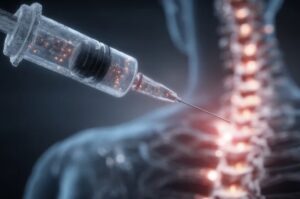
How Microcurrent Helps Scar Tissue
I use a Canadian invented medical device called the Dolphin Neurostim to deliver gentle microcurrent impulses around and directly into scar tissue. These impulses are close to the body’s own bio-electric currents, which is why they can influence healing on a cellular level.
Here’s what happens during treatment:
- RESTORES ELECTRICAL FLOW: Scar tissue can act like an electrical “roadblock.” Microcurrent re-establishes conductivity, normalizing nerve signaling and reducing pain.
- SOFTENS ADHESIONS: By stimulating the tissue, Dolphin therapy encourages reorganization of collagen fibers, improving mobility and pliability.
- REDUCES PAIN & SENSITIVITY: Nerve endings that were trapped or hypersensitive within the scar calm down, often producing immediate relief.
- ENHANCES CIRCULATION: Microcurrent improves microvascular blood flow, which promotes oxygenation and tissue repair.
SIMPLE ANALOGY ➔ Imagine your body’s bio-electric system as a highway network. A scar is like a collapsed bridge—signals can’t get through. Microcurrent helps rebuild that bridge so traffic flows smoothly again.
Early Intervention: Right After Surgery
One of the most exciting aspects of Dolphin Scar Release Therapy is that it can be started almost immediately after surgery (once your medical team has cleared you for safe skin contact). Early treatment has been shown to:
- Reduce post-surgical pain and swelling
- Minimize dense scar formation
- Improve range of motion and healing outcomes
By supporting the body’s natural electrical and fascial systems as healing occurs, the scar often matures more smoothly—leading to fewer long-term issues.
Late Intervention: Decades Later
Even if a scar is decades old, Dolphin therapy can still produce powerful results. Old surgical scars, car accident injuries, C-section scars, or even childhood injuries may continue to influence posture, movement, and pain through both adhesions and electrical disruption.
Patients often report unexpected improvements after treatment—such as relief from chronic low back pain once an abdominal C-section scar is released, or restored shoulder mobility when an old clavicle scar is addressed.
What Else You Can Do at Home
- Gentle movement: Regular mobility exercises keep tissues gliding, but they must be specific to your surgery or injury and guided by your healthcare team. For example, joint replacement surgeries have prescribed mobility protocols, while joint repair surgeries may require immobilization before exercises begin.
- Breath work: Deep, diaphragmatic breathing expands tissues from the inside out, especially helpful after abdominal or chest surgery.
- Hydration: Well-hydrated tissues are more elastic and less prone to stiffness.
- Early care: If you’re recovering from surgery, ask your health team when it’s safe to begin gentle scar work—early guided care often leads to better long-term outcomes.
The Bottom Line
Scar tissue is part of your body’s healing wisdom—but without attention, it can create limitations that last a lifetime. If you’ve had surgery or a significant injury, even years ago, and you’re still experiencing pain or stiffness, your scar tissue may be playing a bigger role than you realize—even influencing how the body communicates electrically.
The good news is that scar tissue doesn’t have to dictate how you move or feel. With Dolphin Neurostim Scar Release Therapy, it’s possible to influence scars right after surgery for better healing outcomes, as well as decades later to resolve long-standing pain patterns. When combined with integrative orthopaedic medical massage, this approach can restore freedom of movement, reduce discomfort, and improve overall quality of life.

by Dena Denali | Sep 2, 2025 | Ask The Expert
What Are Orthopaedic Tests?
Orthopaedic tests are simple but very specific, hands-on assessments. They help identify which structures in the body—muscles, tendons, ligaments, joints, or nerves—might be contributing to your pain or restriction. These tests are not diagnostic in the same way a medical doctor would use imaging or lab tests. However, they give your RMT valuable clinical clues about where the problem is coming from.
For example, a shoulder pain complaint might actually stem from the rotator cuff, a bursa, nerve entrapment, or even a neck muscle. Orthopaedic tests help narrow down the possibilities. Thus, treatment is targeted rather than just a general “rub where it hurts.”
Why They Matter in Massage Therapy
Pain is rarely straightforward. The spot that hurts is often not the true source of the problem. Without assessment, massage can feel good temporarily, but the results may not last. By using orthopaedic tests, your RMT can:
-
Identify whether pain is muscular, joint-related, or nerve-related.
-
Differentiate between acute injury, chronic compensation, or postural strain.
-
Create a treatment plan tailored to your body’s needs rather than guessing.
Think of it as a roadmap—tests show us the fastest, safest route to getting you out of pain and back to function.
A Universal Language Across Healthcare
One of the greatest strengths of orthopaedic testing is its universality. A test performed in an RMT’s office is the sametest recognized and used by physiotherapists, chiropractors, and medical doctors.
That means if your results indicate the need for further investigation—like imaging (X-rays, MRI, ultrasound) or referral to another provider—the findings can be communicated clearly across disciplines. This creates true continuity of care. It ensures you don’t have to start over each time you see a different professional.
Reliability and Clinical Significance
When an orthopaedic test is performed correctly, it has a predictable and repeatable outcome. This consistency is what makes it clinically valid. However, the true value lies in the interpretation of those results.
It’s not just about knowing that, for example, when the hip is flexed to 90 degrees it should internally rotate about 45 degrees and externally about 60 degrees. What matters is understanding what might be limiting that motion—is it muscular tightness, joint restriction, scar tissue, or a neurological factor? This interpretation then directs the next logical test, refines the assessment, and shapes a safe and effective treatment plan.
In other words, orthopaedic testing isn’t just about memorizing numbers or potential outcomes. It requires the therapist to integrate and apply knowledge in real time. They make connections between the test findings, the client’s history, and the overall clinical picture.
This skill can be taught, but it is reinforced and sharpened through ongoing daily use and years of experience. An experienced RMT not only recognizes expected test responses. They also know how to interpret subtle variations—what is significant, what requires further investigation, and what is within normal limits for that individual.
This is why orthopaedic assessment is more than a mechanical process—it’s a dynamic tool that combines standardized testing with professional reasoning, ensuring that each client receives care that is both evidence-informed and personalized.
What to Expect in a Session
Including orthopaedic testing in your session is not complicated or intimidating. Your RMT may ask you to:
-
Resist a gentle push to see how a muscle reacts.
-
Move a joint in a certain direction to test range of motion.
-
Report where you feel discomfort when a certain position is achieved or pressure is applied.
- Or they may perform more complicated maneuvers on your behalf to determine if your results vary whether you are moving the body part versus your RMT
These tests usually take just a few minutes at the start of your appointment but provide information that guides the entire treatment.
The Bottom Line
Orthopaedic assessment is your body’s way of communicating with your RMT. It transforms massage from a feel-good session that lasts a day or so into a targeted, evidence-informed therapy that supports continuity of care, improves outcomes, and helps you move better, recover faster, and stay pain-free longer.

by Dena Denali | Jul 31, 2025 | Ask The Expert, Case Study
Many people assume persistent shoulder pain means a rotator cuff injury, but sometimes the true culprit hides in plain sight: a rotated first rib.
Recently, a patient traveled all the way from Australia to see me in Okotoks, Alberta, after years of unresolved shoulder dysfunction. His story is a perfect example of why assessment-driven, myofascial-based care can reveal what traditional shoulder rehab often misses.
Case Study: 7 Years of Shoulder Pain and Limited Mobility
Patient Profile:
-
35-year-old male — journeyman electrician, strength & conditioning coach for elite athletes, and sensei-level martial artist
-
2010 subscapularis full-thickness tear and shoulder dislocation, surgically repaired with full rehabilitation
-
2018: Martial arts training injury → acute upper trapezius pain, radiating nerve pain into arm and elbow, and sudden inability to raise the arm overhead
-
2022: Pain decreased enough to sleep through the night and regain partial range of motion, but persistent limitations and weakness remained
-
Recent ultrasound radiology report stated thickened subscapularis, supraspinatus and infraspinatus tendons but no evidence of bursitis, but the 2010 surgical repair of subscapularis tendon was noted
Functional Shoulder Assessments of Note:
-
Abduction: 110°
-
Flexion: 150°
-
Internal rotation (with abduction): 50°
-
External rotation (with abduction): 30°
-
Painful and weak in resisted flexion, abduction, and neutral external rotation
Posture & Palpation:
-
Forward head posture and moderate functional thoracic hyperkyphosis
-
Tight anterior musculature (SCM, scalenes, pecs, subscapularis, serratus anterior)
-
Long, inhibited posterior chain (rhomboids, traps, deep neck extensors)
-
Painful first rib costal cartilage at the manubrium joint, A/C joint, and scalene attachment sites
-
Palpable rotated 1st rib in supine
Clinical Insight: Thinking Beyond the Rotator Cuff
At first glance, his history and imaging naturally led previous practitioners to focus on the rotator cuff and surrounding shoulder structures. After all, his surgical history and imaging findings suggested a straightforward shoulder problem. However, despite years of therapy, his pain and functional limitations persisted.
Over time, a pattern began to emerge. Manual therapy and traditional interventions provided brief, temporary relief, but the improvements never lasted. This inconsistency prompted a deeper investigation into the underlying cause of his symptoms.
Through careful orthopaedic assessment and postural analysis, it became clear to me that the true issue extended beyond the rotator cuff itself. A rotated first rib had likely become the primary driver of his ongoing pain, muscular compensation, and restricted mobility—a root problem that traditional shoulder-focused therapy had overlooked.
Treatment Plan
My treatment plan for this case was both integrative and myofascial-focused, combining microcurrent scar release, targeted soft tissue therapy, and ongoing functional reassessment to ensure each session built upon the last.
PHASE 1: Scar and myofascial release
I started by performing microcurrent scar release at the site of his 2010 shoulder surgery, which immediately improved both his range of motion and overall comfort. From there, I used myofascial release and cupping to address tight, overactive anterior muscles including the sternocleidomastoid (SCM), scalenes, pectoralis major and minor, subscapularis, serratus anterior, and latissimus dorsi. To restore muscular balance, I also focused on re-activating the inhibited posterior chain—specifically the rhomboids, mid- and lower trapezius, splenius capitis and cervicis, and semispinalis capitis. This early phase laid the groundwork for improved shoulder and cervical mechanics.
PHASE 2: Functional alignment and joint-specific work
Once the initial restrictions were released, I treated the first rib musculature, including the subclavius, anterior and middle scalenes, and serratus anterior, to restore proper rib and clavicle movement. I also addressed S/C and A/C joint mechanics, as well as key scapular movers such as the levator scapulae, pectoralis minor, latissimus dorsi, and serratus anterior, to improve shoulder stability and overhead motion.
PHASE 3: Neurological and postural reinforcement
To ensure the first rib remained stable and functional gains would hold, I introduced NET (No Extra Time) home care exercises designed to strengthen deep neck flexors, promote scapular retraction, and retrain diaphragmatic breathing patterns. During this stage, we avoided extreme shoulder adduction and clavicular depression to protect the rib’s alignment and allow the surrounding tissues to adapt.
Through this progressive, assessment-driven plan, the treatment addressed not only the symptoms but the root cause of his chronic shoulder dysfunction—creating the conditions for lasting resolution.
Treatment Outcome: Measurable Progress and Key Insights
Over the course of three 90-minute sessions in nine days, we saw both immediate improvements and valuable clinical lessons that shaped the patient’s long-term plan.
During the first session, the rotated first rib was corrected entirely through soft tissue release rather than direct joint mobilization. This approach immediately improved his range of motion and reduced his pain, confirming that the rib alignment was a significant driver of his symptoms. However, in the days following, he experienced a relapse after performing an extreme shoulder adduction movement with cervical rotation, which reproduced his previous discomfort and reduced his shoulder mobility again.
In the second session, we successfully realigned the first rib using the same myofascial release and postural correction approach. This time, the rib remained stable until his third appointment, reinforcing that the treatment approach was effective when he avoided provocative movements.
By the third session, we uncovered a critical piece of the puzzle. While performing soft tissue work on the SCM and scalenes with gentle head rotation, the first rib rotated out of position again. This clearly demonstrated that scalene overactivity combined with cervical rotation was the primary trigger for his rib instability. Once the scalenes were released, the rib returned to a neutral, stable position, confirming both the source of the problem and the path forward for long-term stability.
Through this sequence of sessions, we not only achieved significant pain reduction and improved mobility, but also identified the mechanical triggers behind his chronic first rib rotation. This insight became the foundation for his home care strategy and postural retraining plan, setting him up for continued progress after returning home.
Understanding 1st Rib Rotation
 A rotated first rib is a subtle but significant dysfunction that can mimic rotator cuff injuries or even resemble thoracic outlet syndrome, leading to chronic neck and shoulder pain, restricted movement, and nerve-related symptoms. To fully understand why this occurs, it is important to examine rib anatomy, muscle forces, common triggers, and key symptoms.
Anatomy of the First Rib and Its Joints
From an anatomical perspective, the first rib connects to the spine at the costovertebral and costotransverse joints, which are synovial gliding joints. These joints are inherently stable, but their stability relies heavily on strong ligaments and balanced muscular support rather than deep bony interlocking like the hip or shoulder. Because of this, even small muscle imbalances or postural changes can alter the rib’s position and lead to dysfunction.
How Muscle Forces Influence First Rib Rotation
Muscle forces play a critical role in both stabilizing and rotating the first rib. The anterior and middle scalenes attach directly to the rib, elevating it during inspiration and contributing to anterior or posterior rotation when tension is uneven or chronic. The subclavius muscle, which stabilizes the clavicle, can pull the rib forward when overactive or guarding after injury. Meanwhile, the serratus anterior and upper trapezius indirectly influence rib mechanics through scapular elevation and depression, showing how shoulder function and rib alignment are closely linked.
Common Triggers for First Rib Rotation
Several common triggers can set the stage for first rib dysfunction. Repetitive overhead activity, often seen in electricians, athletes, or martial artists, creates constant loading on the rib and surrounding musculature. Forward head posture and patterns like Upper Cross Syndrome shorten the scalenes and overwork the anterior chain, predisposing the rib to rotation. Additionally, chronic accessory breathing patterns, where scalenes and upper traps dominate over the diaphragm, can perpetuate tension. Trauma, whiplash injuries, or even unresolved postural compensation after surgery can further destabilize the area, and when chronic, the condition can increase the risk of thoracic outlet syndrome.
Symptoms
Symptoms of a rotated first rib often include persistent neck and shoulder tightness, a sense of heaviness or tingling in the arm due to brachial plexus compression, and restricted cervical rotation or side bending. Because these symptoms overlap with more common shoulder injuries, first rib rotation is often overlooked, leading to years of ineffective treatment until the true source is addressed.
Next Steps for This Patient
To ensure long-term success and prevent the first rib from rotating again, the patient left with a structured care planfocusing on myofascial therapy, postural correction, breathing retraining, and progressive strengthening.
Ongoing Myofascial Release for Rib and Shoulder Mechanics
Continued myofascial release will be a key component of his recovery. By replicating the soft tissue techniques that were effective in my clinic, the patient can maintain mobility, prevent scar tissue restrictions, and continue improving shoulder and rib mechanics once back in Australia.
Breathing Retraining to Reduce Scalene Overload
Diaphragmatic breathing is equally essential in the recovery plan. By reducing reliance on the scalenes and upper trapezius as accessory breathing muscles, the patient can offload chronic tension, improve rib stability, and decrease the risk of first rib rotation returning.
Postural and Ergonomic Correction to Address Upper Cross Syndrome
 The next priority is correcting postural imbalances. Years of Upper Cross Syndrome, marked by forward head posture and tight anterior musculature, contributed significantly to the rib dysfunction. Gradually introducing low-load, high-repetition exercises for scapular stabilization and deep neck activation will help the patient restore alignment and prevent recurrence during daily activities.
Strength and Stability Progressions for Lasting Results
Progressive strengthening will follow a phased approach to protect the rib while building long-term stability. Initial isometric exercises encourage rib and scapular stabilization without risking early rotation. Once the rib remains neutral and stable for an extended period, the patient can transition to dynamic exercises that reinforce shoulder and cervical mechanics:
-
Serratus anterior activation: wall slides and push-up plus
-
Lower trapezius and rhomboid engagement: rows and prone Y/T/W exercises
-
Deep neck flexor strengthening: chin tucks and supine nods
By following this integrated plan, the patient can reinforce first rib stability, improve posture, restore functional shoulder movement, and reduce the risk of chronic pain or future relapse.
If Symptoms Persist: Advanced Options for First Rib Instability
Even with a structured treatment plan that includes myofascial release, postural correction, breathing retraining, and progressive strengthening, some patients may still experience recurrent first rib rotation or lingering symptoms. When this occurs, the next step is to reassess the underlying cause of instability and consider advanced interventions.
Diagnostic Imaging to Identify Ligament Laxity
Ultrasound or MRI imaging can reveal whether ligament laxity or microinstability exists at the costovertebral or costotransverse joints. These synovial gliding joints rely heavily on ligamentous and muscular support, so any compromise in these structures can make the first rib more prone to rotation.
Regenerative Injection Therapy for Rib Stability
 If imaging confirms ligament involvement and conservative care has been fully maximized, two regenerative therapy options may help reinforce rib stability:
- PROLOTHERAPY FOR LIGAMENT REINFORCEMENT: Prolotherapy involves injecting a mild irritant solution, often dextrose, into ligament or tendon attachment sites (entheses). The goal is to stimulate collagen production, which tightens and strengthens lax ligaments that contribute to first rib instability.
- PLATELET-RICH PLASMA (PRP) FOR TISSUE HEALING: Platelet-Rich Plasma (PRP) therapy uses a concentrate of the patient’s own platelets injected into targeted soft tissue structures to stimulate healing and tissue repair. While PRP is more commonly used for tendon or enthesis injuries, it can support ligament stability in complex rib cases when combined with functional retraining.
Why Regenerative Injections Are Not a Standalone Solution
It is important to understand that PRP and prolotherapy are not shortcuts. Even if imaging identifies ligamentous laxity, these injections are most successful when combined with postural correction, myofascial therapy, and scapular stabilization exercises. Without addressing muscular compensation and breathing dysfunction, the first rib may continue to rotate despite injections.
Key Takeaway
Ultimately, the path to lasting resolution for chronic first rib rotation is a combination of structural reinforcement and functional retraining. Injections can provide stability at the ligament level, but movement re-education, myofascial therapy, and postural corrections are what make those results sustainable.